Science & Technology, Australia (Commonwealth Union) – 3D bioprinting is an emerging technology that has the potential to revolutionize the field of medicine. It is a process of creating three-dimensional structures using living cells and other biological materials. This technology has the potential to create tissues and organs for transplantation, to create personalized medical devices, and to develop new drugs and therapies.
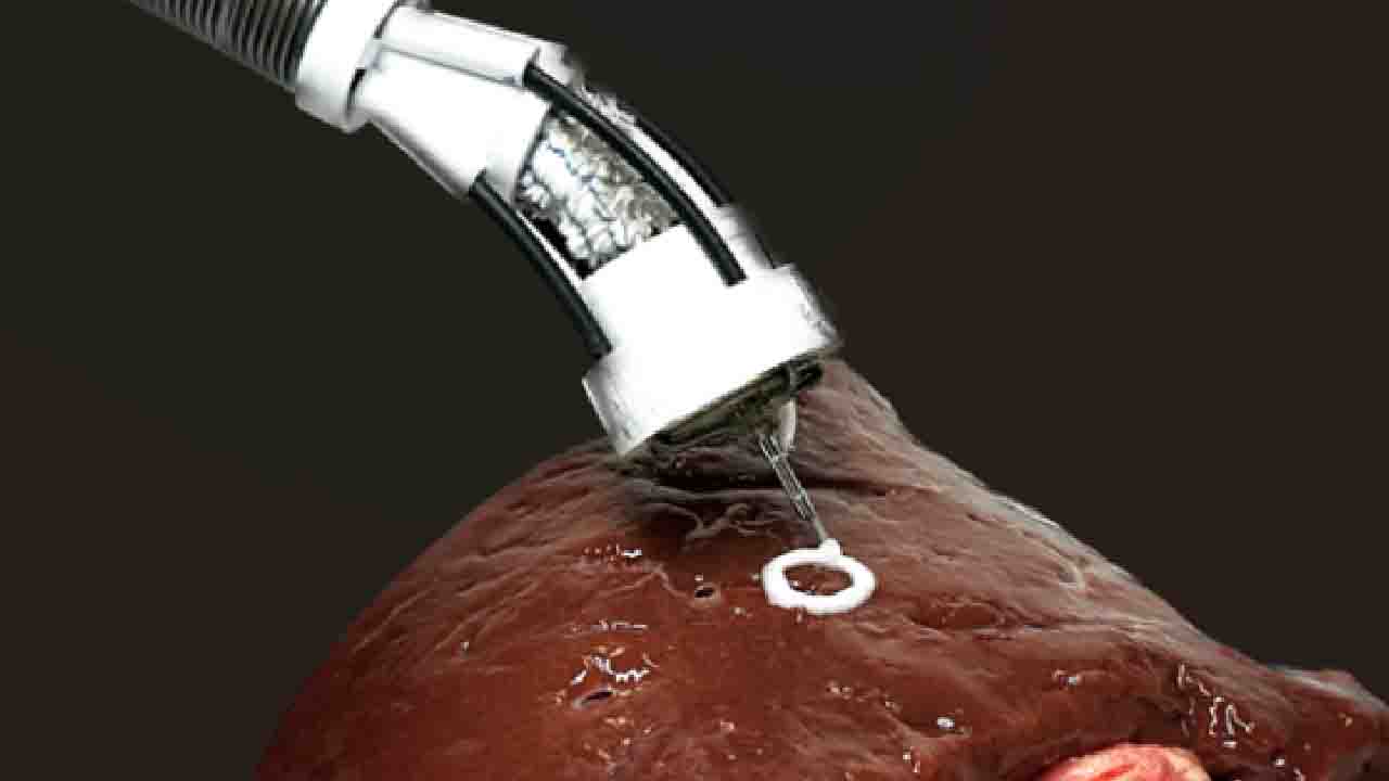
Researchers from the University of New South Wales (UNSW) have shown a model device capable of directly 3D printing living cells onto organs inside and possibly applying it as an all-in-one endoscopic surgical application.
3D bioprinting is a procedure in which biomedical parts are fabricated from a liquid referred to as bioink to form natural tissue-like structures.
Bioprinting is mainly applied in research like tissue engineering and in the formation of new drugs, generally needing the application of bigger 3D printing machines forming cellular structures externally from the living body.
The recent work from UNSW Medical Robotics Lab, led by Dr Thanh Nho Do together with PhD student, Mai Thanh Thai, joining hands with other scientists from UNSW including Scientia Professor Nigel Lovell, Dr Hoang-Phuong Phan, as well as Associate Professor Jelena Rnjak-Kovacina. The findings appeared in Advanced Science.
The research led to a small flexible 3D bioprinter that can be inserted into the body similar to an endoscope and directly transfer multilayered biomaterials to the surface of internal organs along with tissues.
The proof-of-concept device, referred to as F3DB, has a component with increased maneuverability swivel head that ‘prints’ the bioink, joined at the end of a lengthy and flexible snake-like robotic arm, entirely capable of being controlled from the outside.
The researchers indicated that with more development, and possibly in 5 to 7 years, the technology can be utilized by medical professionals accessible in hard-to-reach locations within the body through minor skin incisions or natural orifices.
“Existing 3D bioprinting techniques require biomaterials to be made outside the body and implanting that into a person would usually require large open-field open surgery which increases infection risks,” explained Dr Do, a Scientia Senior Lecturer at the UNSW, Graduate School of Biomedical Engineering (GSBmE) as well as the Tyree Foundation Institute of Health Engineering (IHealthE).
Dr Do further explained that their flexible 3D bioprinter shows that biomaterials can be directly transferred into the target tissue or organs with the lowest invasive approach.
“This system offers the potential for the precise reconstruction of three-dimensional wounds inside the body, such as gastric wall injuries or damage and disease inside the colon.”
“Our prototype is able to 3D print multilayered biomaterials of different sizes and shape through confined and hard-to-reach areas, thanks to its flexible body.”
“Our approach also addresses significant limitations in existing 3D bioprinters such as surface mismatches between 3D printed biomaterials and target tissues/organs as well as structural damage during manual handling, transferring, and transportation process.”
Scientia Professor Nigel Lovell, who is Head of the GSBmE as well as Director of the IHealthE, said “Currently, there are no commercially available devices that can perform in situ 3D bioprinting on internal tissues/organs distanced from the skin surface,” further indicating that certain other proof-of-concept devices were presented, however they have increased rigidity as well as being tricky to utilize in complex and confined places within the body.
The smallest F3DB prototype formed by the team at UNSW has an alike diameter to commercial therapeutic endoscopes, sufficiently minute to be inserted inside the human gastrointestinal tract.
However, the researchers pointed out that they can make it even smaller for future medical applications.
The researchers further plan to implement more features, like an integrated camera along with real-time scanning system that will reconstruct the 3D tomography of the moving tissue from within the body.


