Health, India (Commonwealth Union) – A collaborative effort between the Indian Institute of Science (IISc) and Aster-CMI Hospital has yielded an AI tool capable of identifying the median nerve in ultrasound videos, facilitating the detection of carpal tunnel syndrome (CTS). The research, published in IEEE Transactions on Ultrasonics, Ferroelectrics, and Frequency Control, addresses the challenges posed by ultrasound images in visualizing the median nerve, particularly in areas where its boundaries are less distinct.
Carpal tunnel syndrome arises when the median nerve, extending from the forearm into the hand, experiences compression at the wrist’s carpal tunnel, leading to symptoms like numbness, tingling, or pain. Common among individuals with repetitive hand movements, such as office workers, assembly line employees, and athletes, CTS requires accurate visualization for diagnosis and treatment.
Current medical practices involve using ultrasound to examine the median nerve’s size, shape, and potential abnormalities. However, interpreting ultrasound images can be challenging, especially in regions where other structures obscure the nerve’s boundaries. This complexity is heightened when tracking the nerve in the elbow region. Successful detection of the median nerve is crucial for procedures involving local anesthesia administration or blocking the nerve to alleviate pain, as indicated by researchers of the study.
The research team leveraged a machine learning model based on the transformer architecture, akin to the one powering ChatGPT. Originally designed to simultaneously detect numerous objects in YouTube videos, the model underwent modifications to enhance efficiency, narrowing its focus to track a single object—the median nerve in this instance. Collaborating with Lokesh Bathala, Lead Consultant Neurologist at Aster-CMI Hospital, the team collected and annotated ultrasound videos from both healthy participants and those with CTS to train the model. Once trained, the model demonstrated the capability to accurately segment the median nerve in individual frames of ultrasound videos.
“Imagine a video of an autonomous car. If the car is moving on the road, you want to track the car,” said the corresponding author Phaneendra K Yalavarthy, who is a Professor at CDS. “In the same way, we are able to track the nerve throughout the video.”
The model had the capability to automatically measure the cross-sectional area of the nerve as well, which is applied in identifying CTS. A sonographer makes this measurement manually. “The tool automates this process. It measures the cross-sectional area in real time,” added Bathala. It had the capability to demonstrate the cross-sectional area of the median nerve with over 95 percent preciseness at the wrist region, according to the researchers.
Yalavarthy points out that while numerous machine learning models have been designed for screening CT and MRI scans, there is a notable scarcity of models specifically developed for ultrasound videos, particularly in the context of nerve ultrasound.
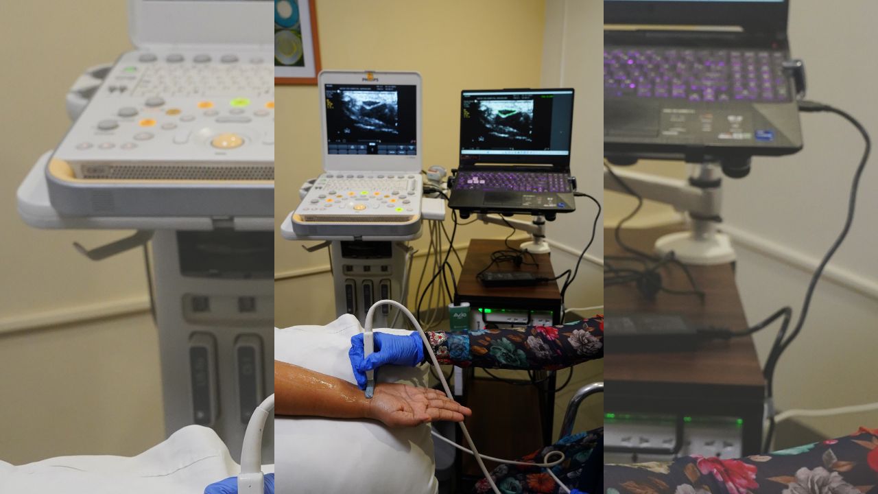
According to Bathala, the initial training of the model focused on a single nerve, and now there are plans to expand its scope to encompass all nerves in the upper and lower limbs. He mentions that the model has undergone a pilot test within the hospital setting, where an ultrasound machine is linked to an additional monitor running the model. This setup allows practitioners to observe the nerve while the software tool simultaneously delineates it, providing real-time performance feedback.
Looking ahead, Bathala emphasizes the intention to collaborate with ultrasound machine manufacturers for the integration of this technology into their systems. A step likely to further enhance the accessibility and applicability of the developed AI tool in clinical settings.
“This kind of tool can assist any doctor. It can reduce the inference time,” he says. “But of course, the final diagnosis will need to be done by the physician.”
The findings are likely to be further enhanced with more research into the area.


