
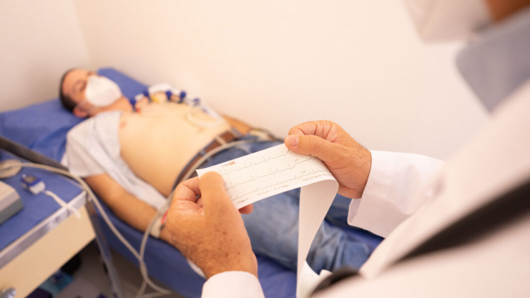

Scientists at the University of California, Davis have tested a new heart device in humans for the first time. The work took place at the National Center for Interventional Biophotonic Technologies, called NCIBT. The device is a thin, bendable catheter. It travels through the arteries and records detailed images of plaque as it goes. Plaque is the fatty buildup that narrows blood vessels. This condition is known as atherosclerosis.
Laura Marcu is the founding director of NCIBT. She says the tool could help doctors understand how heart disease develops. It could also help them plan treatment that fits each patient.
How the device works
The catheter uses two kinds of light-based imaging. The first is fluorescence lifetime imaging, or FLIm. The second is polarization-sensitive optical coherence tomography, or PS-OCT. Together, they show what the plaque is made of and how it is built, all at the same time.
Julien Bec leads engineering and regulatory work in the Marcu Lab. He designed and built both the catheter and the imaging console. He explained that artery walls give off a very weak natural glow. FLIm reads that glow to learn about the chemistry of the tissue. PS-OCT collects light that bounces back from the same spot. This light reveals the fine structure of the wall.
A university lab built this system and ran a patient study. Usually, big companies do that work.
The path to human testing
Before this study, the team tried the device on animals. They also tested it on samples of human heart arteries. This year, the FDA allowed them to study the device in 10 patients as an investigational tool. The team finished imaging the last patient in August.
Why this matters
Heart disease is the top cause of death in the United States. Plaque in the arteries can lead to heart attacks, strokes, and other serious problems.
Doctors use a test called cardiac catheterization to check the heart. They guide a thin tube into a blood vessel. Then they look at blood flow and search for plaque. Current tools, such as standard optical coherence tomography and intravascular ultrasound, show the shape and structure of the vessel. They do not show its chemical makeup. That chemistry matters a great deal, because it affects whether plaque becomes unstable and breaks open.
The new device fills this gap. It captures both structure and chemistry in one pullback. During a pullback, the catheter is slowly drawn back while it records live images.
The catheter moves through the coronary arteries of a beating heart. In just two seconds, it scans 80 millimeters of artery. It can even find plaque before it grows into stenosis. Stenosis is a harmful narrowing that cuts down blood flow.
What doctors say
Jeffrey Southard is an interventional cardiologist at UC Davis. He led the patient tests. He warned that plaque can burst without notice and form blood clots. Those clots may cause a heart attack. He said the catheter should give reliable details about plaque chemistry. Such details may point to inflammation and a risk of rupture or erosion. More research will guide treatment choices. Postdoctoral researcher Xiangnan Zhou ran the system in the cath lab.
Looking ahead
Many patients with plaque need stents to hold arteries open. Where doctors place the stents matters. So does the choice of drugs. These choices affect how well treatment works over the long term.
The team now plans to enlarge the study and include more patients. Marcu hopes cath labs will one day use the device to assess plaque makeup. That knowledge could guide how doctors treat patients and place stents.